|
Major research areas of our studies concern:
1. Concentration of vestigial elements
and degree of degeneracy of man's intestine cells
(pl version)
Background
back In collisions of heavy ions
with target atoms the strong Coulomb field of one of the collision partners' can
cause simultaneous ejection of several electrons of the second one. This process
results in a reduction of the nuclear charge screening and increases the binding
energy of remaining electrons [1]. Consequently, the energies of x-rays emitted from
such multiply ionized atoms are shifted with respect to the corresponding x-ray energies
of singly ionized atoms and reflect the actual configuration of electrons during x-ray emission.
Finally, as a result of the multiple ionization, instead of a single-hole x-ray transition
called the diagram line, the structure of x-ray satellites appears.
Collision processes have been studied extensively for many years but these studies were focused
mainly on single or multiple K-, L-, M-shell ionization occurring in the target atoms. The satellite
structure of K and L x-ray lines of the target atoms, observed mainly with high resolution spectrometers
[2-5], was interpreted as the result of additional vacancies in outer shells of the atom. However, more
complicated processes are experienced by a projectile as a second partner of the ion-atom collision.
During the first collision K-vacancy and/or multiple L-, M-, N- or higher shell vacancies can be produced.
Further, the ion can collide with the other target atoms and capture or loss of electrons can occur before
its original vacancies are filled by outer electrons. The competition between ionization, excitation, electron
capture, electron loss and decay processes leads to the establishment of an electron-vacancy equilibration
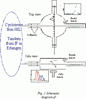
in different shells of the moving projectile. Sulphur ion beams with incident energies of 9.6, 16.0, 22.4, 32.0 MeV and with initial charge states q=4+, 6+ were obtained from the tandem accelerator at the Institute of Physics of the Erlangen Nürnberg University. Other energies 65, 79, 99 and 122 MeV and with incident charges of 13+ and 14+ were obtained from the U-200P cyclotron at the Heavy Ion Laboratory of Warsaw University. A schematic diagram of the experimental arrangement is shown in figure 1. Two collimators located at 24 and 38 cm in front of the target are used to define 2 mm in diameter beam spot on the target. Self-supporting carbon foils with effective thickness of 15-210 µg/cm2, were positioned in the target holder at the center of vacuum chamber at an angle of 25o to the direction of the beam. The geometry of the experimental arrangement used in this work means that the detector should register x-rays emitted by projectile inside the target as well as from distance up to 1.2 cm behind the target, so during such experiments x-rays with lifetimes up to 10-12 sec are registered.
The carbon targets were prepared by
vacuum evaporation and their absolute thickness was determined in the separate measurements
of energy loss of 5.48 MeV a particles from 241Am source. The stopping power values for a
particles in carbon needed to calculate the foil
thickness were obtained from Biersack and Maggamark [40] and the final
target thickness was calculated by the computer code TRIM [41]. Additionally
these thicknesses were checked using elastic scattered 2.0 MeV 4He+ ions from the
Van de Graaff accelerator. Absolute target thicknesses were determined with the accuracy
of about ą 4%. The targets could be considered as `thin' because the ions passing through
the foil did not lose energy appreciably (DE was less than 0.15Eo for the thickest target
and for the lowest ion energy and decreased rapidly up to 0.01Eo for the highest projectile energy).
The effective ion energy was further used. The self-absorption of the measured x-rays in a target
is also small (less than 4%). Independent measurements of target thickness enabled absolute normalization
of the x-rays intensity on the incident number of projectiles obtained from elastically scattered sulphur
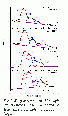
ions detected in a silicon surface-barrier detector located inside the chamber at 12.5o to the beam direction. Typical x-ray spectra recorded by Si(Li) detector for sulphur ions passing with energies of 16, 22.4, 79 and 122 MeV through a carbon target are presented in Fig. 2.
The origin of all recorded peaks is described in detail in our previous paper [30]. The resolved
KaS1,2,
KbS1,3 satellite and
Kah1,2,
Kbh1,3 hypersatellite
peaks are the results of the overlapped contributions corresponding
to transitions of the following types: [1] P. H. Mokler and F. Folkmann, in: I. A. Sellin (Ed.), Structure and Collisions of Ions and Atoms, Springer Verlag, Berlin, p. 201 (1978). [2] J. McWherter, J. Bolger, C. F. Moore, and P. Richard, Z. Phys. 263, 283 (1973). [3] R. L. Kaufman, J. H. McGuire, P. Richard, and C. F. More, Phys. Rev. A8, 1233 (1973). [4] R. L. Watson, F. E. Jenson, and T. Chiao, Phys. Rev. A10, 1230 (1974). [5] P. Rymuza, Z. Sujkowski, M. Carlen, J-Cl. Dousse, M. Gasser, J. Kern, B. Penry, and Ch. Rheme, Z. Phys. D14, 37 (1989). [6] H. D. Betz, F. Bell, H. Panke, G. Kalkoffen, M. Welz, and D. Evers, Phys. Rev. Lett. 33, 807 (1974). [7] F. Hopkins, Phys. Rev. Lett. 35, 270 (1975). [8] K. O. Groeneveld, B. Kolb, J. Schader, and K. D. Sevier, Z. Phys. A277, 13 (1976). [9] T. J. Gray, P. Richard, K. A. Jamison, and J. M. Hall, Phys. Rev. A14, 1333 (1976). [10] F. Hopkins, J. Sokolov, and A. Little, Phys. Rev. A 15, 588 (1977). [11] R. K. Gardner, T. J. Gray, P. Richard, C. Schmiedekamp, K. A. Jamison, and J. M. Hall, Phys. Rev. A15, 2202 (1977). [12] J. A. Tanis, W. W. Jacobs, and S. M. Shafroth, Phys. Rev. A22, 483 (1980). [13] B. B. Dhal, H. C. Padhi, K. G. Prasad, P. N. Tandon, and M. Polasik, J. Phys. B: 31, 1225 (1998). [14] P. H. Mokler, Phys. Rev. Lett. 26, 811 (1971). [15] C. W. Woods, F. Hopkins, R. L. Kauffman, D. O. Elliott, K. A. Jamison, and P. Richard, Phys. Rev. Lett. 31, 1 (1973). [16] R. L. Watson, J. R. White, and F. E. Jenson, Phys. Lett. A67, 269 (1978). [17] R. L. Watson, J. R. White, A. Langenberg, R. A. Kenefick, and C. C. Bahr, Phys. Rev. A22, 582 (1980). [18] Y. Awaya, T. Kambara, M. Kase, H. Shibata, H. Kumagai, K. Fujima, J. Urakawa, T. Matsuo, and J. Takahashi, Nucl. Instrum. And Methods Phy. Res. B 10/11, 53 (1985). [19] K. Shima, K. Umetani, and T. Mikumo, Nucl. Instrum. and Methods 194, 353 (1982). [20] H. J. Hay, L. F. Pender, and P. B. Treacy, Aust. J. Phys. 34, 155 (1981). [21] H. J. Hay, L. F. Pender, and P. B. Treacy, Nucl. Instrum. And Methods 194, 349 (1982). [22] Y. Zou, Y. Awaya, C. P. Bhalla, T. Kambara, Y. Kanai, M. Oura, Y. Nakai, K. Ando, A. Hitachi, and S. Kravis. Phys. Rev. A 51, 3790 (1995). [23] T. Mizogawa, Y. Awaya, T. Kambara, Y. Kanai, M. Kase, H. Kumagai, P. H. Mokler, and K. Shima, Phys. Rev. A 42, 1275 (1990). [24] H. F. Beyer, H. J. Kluge, and V. P. Shevelko, X-Ray Radiation of Highly Charged Ions, Springer Series on Atoms and Plasmas, Springer-Verlag, Berlin Heidelberg, New York (1997). [25] M. Pajek, D. Bana, J. Braziewicz, U. Majewska, J. Semaniak, T. Czyżewski, M. Jaskóła, W. Kretshmer, T. Mukoyama, D. Trautmann, and G. Lapicki, AIP Conf. Proc. 475, 32 (1999). [26] D. Bana, M. Pajek, J. Semaniak, J. Braziewicz, A. Kubala-Kuku, U. Majewska, T. Czyżewski, M. Jaskóła, W. Kretschmer, T. Mukoyama, and D. Trautmann, Nucl. Instrum. and Methods Phy. Res. B 195, 233 (2002). [27] D. Bana, J. Braziewicz, U. Majewska, M. Pajek, J. Semaniak, T. Czyżewski, M. Jaskóła, W. Kretschmer, and T. Mukoyama, Nucl. Instrum. and Methods Phy. Res. B 154, 247 (1999). [28] D. Bana, J. Braziewicz, A. Kubala-Kuku, U. Majewska, M. Pajek, J. Semaniak, T. Czyżewski, M. Jaskóła, W. Kretschmer, and T. Mukoyama, Nucl. Instrum. and Methods Phy. Res. B 164-165, 344 (2000). [29] D. Bana, J. Braziewicz, U. Majewska, M. Pajek, J. Semaniak, T. Czyżewski, M. Jaskóła, W. Kretschmer, T. Mukoyama, and D. Trautmann, J. Phys. B 33, L793 (2000). [30] U. Majewska, K. Słabkowska, M. Polasik, J. Braziewicz, D. Bana, T. Czyżewski, I. Fijał, M. Jaskóła, A. Korman, and S. Chojnacki, J. Phys. B 35, 1941 (2002). [31] R. L. Watson, J. M. Blackadar, and V. Horvat, Phys. Rev. A 60, 2959 (1999). [32] R. L. Watson, V. Horvat, J. M. Blackadar, and K. E. Zaharakis, Phys. Rev. A 62, 052709 (2000). [33] H. Panke, F. Bell, H. -D. Betz, H. Stehling, E. Spindler, and R. Laubert, Phys. Lett. 53A, 457 (1975). [34] H. Panke, F. Bell, H. D. Betz, and H. Stehling, Nucl. Instrum. and Methods 132, 25 (1976). [35] J. A. Tanis and S. M. Shafroth, Phys. Rev Lett. 40, 1174 (1978). [36] U. Scharfer, C. Henrichs, J. D. Fox, P. Von Brentano, L. Degener, J. C. Sens, and A. Pape, Nucl. Instrum. and Methods 146, 573 (1977). [37] T. J. Gray, C. L. Cocke, and R. K. Gardner, Phys. Rev. A 16, 1907 (1977). [38] C. L. Cocke, S. L. Varghese, and B. Curnutte, Phys. Rev. A 15, 874 (1977). [39] S. K. Allison, Rev. Mod. Phys. 30, 1137 (1958). [40] J. P. Biersack and L. G. Maggmark, Nucl. Instrum. and Methods 174, 257 (1980). [41] J. Ziegler, http:\\ www.srim.org [42] M. Pajek, A. P. Kobzev, R. Sandrik, R. A. Ilkhamov, and S. A. Khusmorodov, Nucl. Instrum. and Methods Phy. Res. B 42, 346 (1989). [43] U. Majewska, J. Braziewicz, M. Polasik, K. Słabkowska, I. Fijał, M. Jaskóła, A. Korman, S. Chojnacki, and W. Kretschmer, Nucl. Instrum. and Methods Phy. Res. B 205, 799 (2003). Results 1. A new theoretical model based on the results of the single-configuration DF calculations (and using equilibrium charge state distribution and fluorescence yields) has been proposed in order to estimate the role of various types of electronic configurations and to evaluate the average population of different shells in fast sulphur ions passing through the carbon foils from low resolution K x-ray spectra. [U. Majewska et al., Configurations of highly ionized fast sulphur projectiles passing through a crbon foil evaluated from low-resolution K x-ray spectra. J. Phys. B: At. Mol. Opt. Phys. 35 (2002) 1941]. 2. A new interpretation of low resolution K x-ray spectra parameters of highly ionized sulphur projectiles passing through the carbon foils has been proposed using theoretical model based on the single-configuration Dirac-Fock calculations, equilibrium charge state distribution and fluorescence yields for multiply ionized sulphur ions, the population of L and M shells (in the case of 9.6 - 32.0 MeV beam energy), the probability of K-hole creation and the average population of L-shell, 3p and 4p subshells (in the case of 65 - 122 MeV) of sulphur ions have been estimated. Additionally, the lifetimes of discussed highly excited states of sulphur ions have been obtained from the studying of dynamics of formation of K-hole fractions of sulphur projectiles projectiles inside a carbon foil. [U. Majewska, et al., Highly excited states of sulphur projectiles inside a carbon target, Nucl. Instrum. and Meth. in Phys. Res. B205 (2003) 799]. 3. The dependence of the satellite and hypersatellite sulphur K x-ray production cross sections on the target thickness has been examined, for the first time separately for each line recorded in spectra of characteristic x-rays of the projectiles at energy range of 65-122 MeV. The three component model, which expresses the probability of an ion charge-changing collisions by the cross sections sij was used. For each sulphur projectile energy the values of the K-shell vacancy production cross sections and the K-shell vacancy loss cross sections (independently for the electron capture and for the radiative and Auger decays) have been fitted. The obtained experimental values of all sij cross sections have been used later to determine the dynamics of formation of the K-vacancy fractions of the sulphur projectiles passing through a carbon foil. [J. Braziewicz, et al., Dynamics of formation of K-hole fractions of sulphur projectiles inside a carbon foil, Phys. Rev. A69 (2004) 062705] 4. It has been shown that the energies of K X-ray lines emitted from sulphur projectiles are very sensitive to the degree of ionization of their L and M shells. Therefore probably for the first time, the projectile low-resolution K X-ray spectra have been used for evaluation of a mean equilibrium charge, |
|
|
Last modified:
2018-06-12 |
Webmaster:
Dr.Sc.
Janusz Braziewicz
|